Carbon monoxide (CO) poisoning, whether accidental or suicidal, is the most common cause of sequelae and deaths caused by inhalation poisoning worldwide. It is thought to be responsible for approximately 31% of all poisonings in our country. In our country, CO poisonings are mostly seen in winter months and in windy weather. Accidental deaths can be seen in winter months and suicidal deaths can be seen at any time of the year.
Carbon monoxide affects cellular functions together with endogenously produced CO and nitric oxide during hemoglobin catabolism in humans. Endogenously produced CO binds to 0.4-0.7% of human hemoglobin and forms carboxyhemoglobin (COHb). This acts as a neurotransmitter. However, COHb has been found to be 1-3% as a result of environmental exposure and 6-15% in smokers. Exogenous CO is formed by incomplete combustion of carbonaceous compounds such as coal, oil, fertilizer, dung, natural gas. Exogenous sources include exhaust gases from motor vehicles, heaters, methylene chlorides and fires. In closed garages, lethal levels of COHb can be reached within 10 minutes.
Carbon monoxide; CO is a colorless, odorless, non-irritating, toxic gas that is easily absorbed by the lungs. CO is mainly eliminated unchanged by the lungs. Less than 1% of it is converted to carbon dioxide, 10-15% is bound to proteins such as myoglobin and cytochrome-c oxidase, and less than 1% is in solution. As is known, CO toxicity has direct damaging effects on tissue hypoxia and cellular level. CO competes with oxygen for oxygen binding and binds to hemoglobin 200-250 times more than oxygen. This change causes a decrease in oxygen carrying capacity, impaired oxygen release at the tissue level and cellular hypoxia. Levels of small amounts of free CO dissolved in the blood play an important role. The direct effect of CO on cells suggests that the impairment of oxygen transport capacity is also important. CO disrupts cellular respiratory function by reversibly binding to intracellular oxygen carrier heme proteins and causes mitochondrial disruption and tissue damage, especially in brain and heart cells with high energy needs. CO has a very high affinity for myoglobin. Binding of CO to cardiac myoglobin causes myocardial depression, hypotension and arrhythmias. After hypoxia in tissues, oxygenation damage begins to occur in the central nervous system. Hypoxia causes advanced mitochondrial disruption, capillary leakage, lococyte accumulation and cell destruction in the brain. As a result, reversible demyelination occurs in the brain. Basal ganglia are the most frequently affected areas due to high oxygen consumption.
In pregnant women, exposure to carbon monoxide can have serious harmful effects on the fetus. Because the fetus is more sensitive to CO, COHb levels exceed maternal levels. In the fetus, the CO half-life is prolonged, oxygen release to fetal tissues is reduced and severe tissue hypoxia occurs.
Clinically, the severity of poisoning depends on the CO concentration and the duration of exposure. Children, the elderly, people with heart disease, anemia, lung disease and pregnant women are at greater risk. CO poisoning can be acute and chronic. Clinical findings differ accordingly. Headache, weakness, dizziness, drowsiness, drowsiness, difficulty in thinking, chest pain, palpitations, nausea and diarrhea may be observed in low dose and prolonged exposure. Acute poisonings are diagnosed more easily than chronic ones. Neurologic and cardiologic findings may be prominent in acute poisoning. Loss of consciousness, impaired balance and gait, hypotension, heart attacks, rhythm irregularities and metabolic acidosis may occur. Loss of consciousness is the most common neurological finding. In addition, nonspecific findings such as syncope, seizures, memory loss, dizziness, cognitive disorders, balance and gait disturbances, tachycardia, tachypnea, headache, nausea, vomiting, lethargy are common and may be confused with viral infections. In CO poisoning, COHb value rises very high in long-term exposures. As a result, both cardiac and neurologic late findings occur. Severe headache, Parkinson’s disease symptoms, heart attacks, hypothyroidism, more often hypertroidism, and irregularities in reproductive hormones in men and women may be observed.
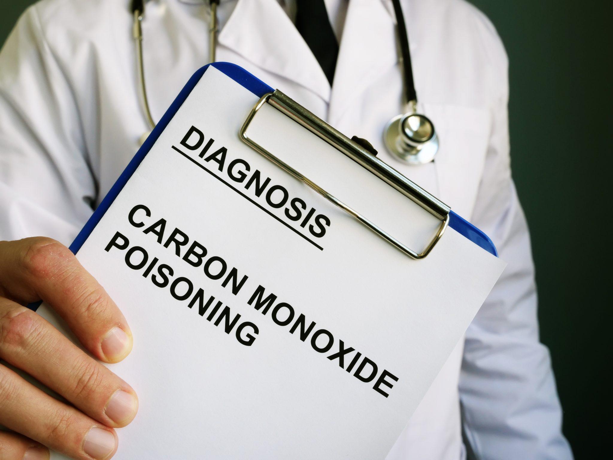
Diagnosis; the most important diagnostic tool is the physician’s suspicion of CO poisoning. A good history should be taken in patients with suspected CO poisoning. Stoves, heaters, closed garages etc. at home and workplace should be questioned. Especially the cause of the event, duration of exposure to CO, loss of consciousness even if transient, chest pain, other poisoning possibilities such as cyanide or drugs should be investigated. If there is suspicion, COHb levels in venous or arterial blood gas should be checked. The most important finding in the diagnosis is the measurement of COHb level in the blood. COHb levels lower than 10% may not be related to the patient’s symptoms, but COHb levels higher than 10% may be diagnostic. While findings such as headache, mild dyspnea and gastroenteritis are observed at COHb levels of 10-30%, COHb levels of 40% and above may cause severe findings such as coma, convulsion and cardiac arrest. Metabolic acidosis in blood gas is considered as severe intoxication. In addition, lactate level is also important.
Patients should be evaluated for cognitive functions, loss of strength, balance, gait and speech in neurologic examination. In the emergency room, hypoglycemia may be confused with CO poisoning. In case of a change in consciousness, brain computed tomography should be performed. Lesions at the level of globus pallidum are most commonly seen in CO poisoning. Magnetic resonance imaging should also be performed for white matter abnormalities. Delayed neuropsychiatric disease may occur weeks (between 3-240 days) after the acute intoxication symptoms resolve. This syndrome is estimated to occur in 10-30% of patients. In these patients, disturbances of consciousness, personality changes such as aggression, violence, impulsiveness, irritability, parkinsonism, urinary incontinence, dementia and psychosis have been reported. In delayed neuropsychiatric syndromes, recovery is seen in 50-75% within 1 year.
Electrocardiography should be performed in all patients with carbon monoxide poisoning. Ischemic changes, ST depression, ST elevation may be seen despite normal coronary arteries. If ST elevation is present in CO poisoning, thrombolysis is probably not appropriate because the cause is myocardial toxicity rather than thrombotic occlusion of epicardial coronary arteries. ECG monitoring should be performed to monitor arrhythmia.

Treatment; carbon monoxide reversibly binds to hemoglobin and tissue proteins faster than oxygen. The basis of treatment in CO poisoning is the removal of CO. Although the half-life of COHb is variable, it is 4-6 hours. Therefore, oxygen should be given to all patients with CO poisoning until COHb drops below 5%. Hyperbaric oxygen therapy is very useful in severe cases. Since lactic acidosis facilitates tissue oxygen diffusion, it should not be corrected unless pH<7.15. In addition to shortening the COHb half-life, hyperbaric oxygen therapy has positive effects such as decreasing oxygen free radical production and levels, inhibition of lipid peroxidation, correction of impaired mitochondrial functions, and correction of impaired platelet adhesion to capillaries. Currently accepted Hyperbaric oxygen therapy includes loss of consciousness, neurological or cognitive abnormalities, cardiac ischemia findings on electrocardiography, COHb levels higher than 20% and pregnancy.
Prevention; Public education on the dangers of CO is important in reducing sequelae and deaths from CO poisoning. This education should be emphasized through the media during the winter months. Regular maintenance of heating systems in homes and workplaces, adequate ventilation and cleaning of chimneys can prevent CO poisoning in homes and workplaces. CO sensors can warn against acute and chronic poisoning. Catalytic converters can reduce the CO content in vehicle exhaust gases, thereby reducing the severity of CO exposure.

Doç.Dr. Abuzer COŞKUN
Bağcılar Training and Research Hospital
Emergency Medicine Clinic
Education Officer